
Fig. 6-S2
- ID
- ZDB-FIG-180821-53
- Publication
- Fukui et al., 2018 - Hippo signaling determines the number of venous pole cells that originate from the anterior lateral plate mesoderm in zebrafish
- Other Figures
- All Figure Page
- Back to All Figure Page
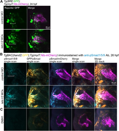
Depletion of Lats1/2 leads to an activation of Bmp-Smad signaling. (A) Confocal 3D-stack images (at 24 hpf) of Tg(BRE:GFP);Tg(myl7:Nls-mCherry) embryos injected with control MO (n = 11) and lats1/2 MOs (n = 12). Square brackets indicate the region in which BRE-dependent GFP-positive myl7-promoter-active cells are present in the venous pole. (B) Confocal images (at 26 hpf) of TgBAC(hand2:GFP);Tg(myl7:Nls-mCherry) embryos injected with control MO (n = 16) or lats1/2 MOs (n = 14). Square brackets indicate the phosphorylated Smad1/5/9-positive myl7-promoter-active cells in the venous pole. Confocal 3D-stack images (the most-right panels) and single scan images (left four panels). (C) Images of TgBAC(hand2:GFP);Tg(myl7:Nls-mCherry) embryos treated with DMH1 (10 μM) from 14 hpf to 26 hpf (n = 6) and immunostained with the anti-pSmad1/5/9 Ab. Both pSmad1/5/9-positive and hand2-promoter-active cells in the venous pole are decreased in the embryos treated with DMH1. (B,C) Dorsal view, anterior to the top. The confocal 3D-stack images and single-scan images are a set of representative images from at least three independent experiments. |