
Fig. 2-S2
- ID
- ZDB-FIG-180821-42
- Publication
- Fukui et al., 2018 - Hippo signaling determines the number of venous pole cells that originate from the anterior lateral plate mesoderm in zebrafish
- Other Figures
- All Figure Page
- Back to All Figure Page
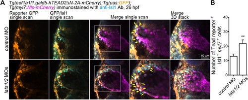
Depletion of Lats1/2 leads to an increase in the number of both Tead-reporter and Isl1-positive SHF cells in the venous pole. (A) Confocal images (at 26 hpf) of Tg(eef1a1l1:galdb-hTEAD2ΔN-2A-mCherry);Tg(uas:GFP);Tg(myl7:Nls-mCherry) embryos injected with the MOs indicated on the left and immunostained with the anti-Isl1 Ab. The boxed regions in the center panels are enlarged in the next right panels. Tead reporter-dependent GFP-positive cells in the Isl1-positive myl7-promoter-active cells (yellow arrows) are observed in the venous pole. Confocal 3D-stack images (the panels furthest to the right) and single scan images (left four columns). Dorsal view, anterior to the top. (B) Quantitative analyses of the numbers of Isl1-positive myl7-promoter-active cells that are positive for Tead-reporter-dependent GFP in the venous pole of (A) (n = 5). **p < 0.01. The confocal 3D-stack images and single-scan images are a set of representative images from three independent experiments. |