
Fig. S2
- ID
- ZDB-FIG-180516-22
- Publication
- Ojeda Naharros et al., 2017 - Loss-of-function of the ciliopathy protein Cc2d2a disorganizes the vesicle fusion machinery at the periciliary membrane and indirectly affects Rab8-trafficking in zebrafish photoreceptors
- Other Figures
- All Figure Page
- Back to All Figure Page
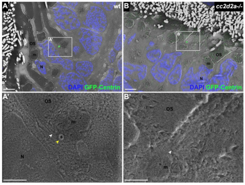
BB docking occurs normally in cc2d2a-/- PRs. 5 dpf CLEM sections of transgenic tg(tacp:GFP-hCentrin) wild-type (wt) (A) and cc2d2a-/- (B) fish expressing GFP-tagged centrin and counterstained with DAPI (blue, nuclei). GFP-Centrin-labeled basal bodies (BBs) (green) localize at the apical membrane of both wt and mutant animals. (A’) BB is docked right below the outer segment (white arrowhead), apical to the daughter centriole (yellow arrowhead) in wt. (B’) BB (white arrowhead) is localized correctly in cc2d2a-/- PRs even when the OSs appear dysmorphic and disorganized. Scale bars: 4 μm in A-B and 2 μm in A’-B’. OS outer segment, m mitochondria, N nucleus, wt wild-type. |