
Fig. S1
- ID
- ZDB-FIG-180516-21
- Publication
- Ojeda Naharros et al., 2017 - Loss-of-function of the ciliopathy protein Cc2d2a disorganizes the vesicle fusion machinery at the periciliary membrane and indirectly affects Rab8-trafficking in zebrafish photoreceptors
- Other Figures
- All Figure Page
- Back to All Figure Page
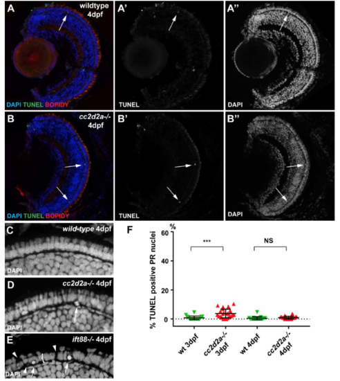
cc2d2a-/- retinae do not undergo degeneration at early developmental stages. (A-B”) TUNEL assay on 4 dpf retinal cryosections of wild-type (A-A”) and cc2d2a mutants (B-B”). Note the limited number of TUNEL positive cells (B’ and green in B) in mutant retinae. Membranes are counterstained with BODIPY (red in A-B) and nuclei with DAPI (A”-B” and blue in A-B). Also note the normal organisation of mutant retina, including the photoreceptor (PR) cell layer, visible with DAPI in (B”) compared to wild-type in (A”). (C-E) Higher magnification views of the PR cell layer in wild-type (C), cc2d2a mutant (D) and ift88 mutant (E) 4 dpf larvae. Note the normal nuclear morphology in cc2d2a-/- PRs compared to wild-type and compared to the degenerating ift88 retina which displays rounded nuclei (arrows in E), gaps (arrowheads in E) and a globally thinned PR cell layer (bracket in E). (F) Quantification of TUNEL positive cells in wild-type (green inverted triangles) and in cc2d2a mutant (red triangles) at 3 and 4 dpf. While the amount of cell death is statistically significantly increased at 3 dpf in mutant compared to wild-type, it remains minimal (on average 4.6% of evaluated nuclei are TUNEL positive in mutants, compared to 0.8% in wild-type). At 4 dpf, no increase in cell death is observed in cc2d2a mutant retinae compared to wild-type. NS non significant, *** p<0.001, t-test, n>20 animals for each condition. Quantification was performed on confocal stacks of identical dimensions in wild-type and mutant. |