
Fig. 1
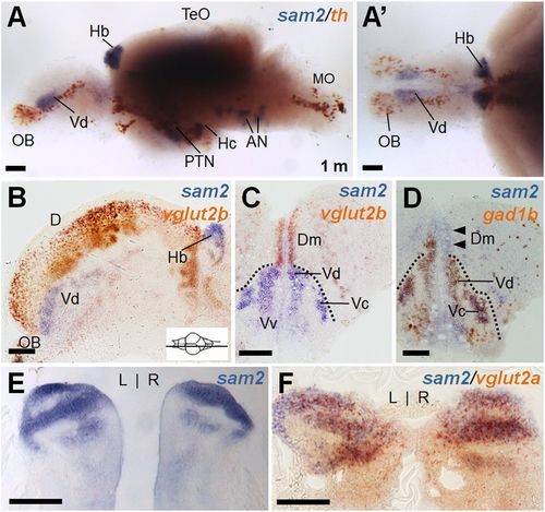
Characterization of sam2-expressing cells in the adult zebrafish brain. (A and A′) Whole-mount two color in situ hybridization of a dissected zebrafish brain with sam2 and a dopaminergic neuron marker, th. Anterior is to the left; lateral (A) and dorsal view (A′). The sam2 expression region did not overlap with th+ neurons. Prominent sam2 expression is seen in the Vd and Hb as well as hypothalamic regions. (B–D) Section images of brain hybridized with sam2/vglut2b (B, sagittal section; C, cross-section) or sam2/gad1b (D). (E and F) Cross-section of Hb region stained with sam2 alone (E) or sam2/vglut2a (F) probes. AN, auditory nerve; D, area dorsalis telencephlali; Dc, central zone of area dorsalis telencephali; MO, medulla oblongata; OB, olfactory bulb; TeO, optic tectum; Vv, ventral nucleus of area ventral telencephali. (Scale bars, 100 µm.) |