
Fig. S4
- ID
- ZDB-FIG-170828-1
- Publication
- Wehner et al., 2017 - Wnt signaling controls pro-regenerative Collagen XII in functional spinal cord regeneration in zebrafish
- Other Figures
- All Figure Page
- Back to All Figure Page
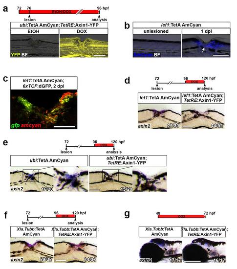
Utilizing the TetON system for cell type-specific Wnt/β-catenin pathway manipulation. (a) The TetON system allows for inducible transgene expression. YFP fluorescence (TetResponder transgene) is robustly induced in DOX-treated but not EtOH-treated ubi:TetA AmCyan;TetRE:Axin1-YFP double transgenic animals. (b) Consistent with Wnt/β-catenin pathway activation, AmCyan fluorescence is undetectable in unlesioned lef1:TetA AmCyan transgenic animals and upregulated after a lesion (arrow). (c) gfp mRNA (green) and amcyan mRNA (red) are co-expressed in lesioned 6xTCF:dGFP;lef1:TetA AmCyan double transgenic animals, indicating that lef1 regulatory elements drive gene expression in Wnt-responding cells in the lesion site. (d) DOX treatment of lef1:TetA AmCyan;TetRE:Axin1-YFP double transgenic animals (but not single transgenic control animals) interferes with axin2 expression in non-neural lesion site cells. (e) Ubiquitous axin1 overexpression through DOX treatment of ubi:TetA AmCyan; TetRE:Axin1-YFP double transgenic animals, interferes with axin2 expression in non-neural lesion site cells. (f) axin1 overexpression specifically in neurons through DOX treatment of Xla.Tubb:TetA AmCyan;TetRE:Axin1-YFP double transgenic animals does not reduce axin2 expression in non-neuronal lesion site cells. (g) DOX treatment of Xla.Tubb:TetA AmCyan;TetRE:Axin1-YFP double transgenic animals (but not single transgenic control animals) reduced axin2 expression in a constitutively Wnt-responsive domain in the brain (arrows), indicating that the Xla.Tubb:TetA AmCyan TetActivator line drives functionally relevant axin1 levels in neurons. (a-g) Views are lateral (dorsal is up, rostral is left). BF: brightfield. Scale bars: whole mounts, 200 μm (g), 100 μm (a-b, d-f) and 25 μm (c). |