
Fig. 5
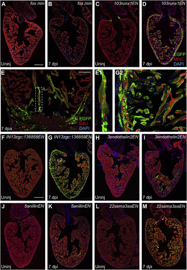
Regeneration Enhancers Direct Gene Expression in CMs after Injury (A and B) Uninjured adult hearts from transgenic reporter lines containing the c-fos minimal promoter alone do not express EGFP whether uninjured (Uninj) or after induced ablation of CMs (7 dpi). Scale bar, 200 μm. (C and D) Uninjured 103runx1ENfos:EGFP animals express EGFP in CMs adjacent to valves. Ablation injury induced EGFP throughout regenerating CMs, most obvious in the compact layer. Scale bar, 200 μm. (E) EGFP fluorescence is induced at 103runx1ENfos:EGFP injury sites at 7 days after apical resection (7 dpa). EGFP-positive CMs at the site of injury in 103runx1ENfos:EGFP hearts incorporate EdU (red). Scale bar, 200 μm. (E1) High-magnification view of box 1 in (E). (F and G) IN13unkENfos:EGFP hearts have low myocardial EGFP fluorescence that is enhanced by ablation injury. Scale bar, 200 μm. (G2) High-magnification view of box 2 in (G). (H and I) 3endothelin2ENfos:EGFP animals express EGFP in CMs near the ventricular lumen and occasionally in the compact layer. Expression increases in regenerating CMs at 7 dpi. (J and K) 5anillinENfos:EGFP animals display EGFP sporadically in uninjured hearts. Expression increases in regenerating CMs after induced ablation of CMs. (L and M) 22sema3aaENfos:EGFP zebrafish show induced expression throughout CMs only after injury. An antibody against myosin heavy chain (MHC, red) was used to stain cardiac muscle. See also Figure S5. |
| Gene: | |
|---|---|
| Fish: | |
| Conditions: | |
| Anatomical Terms: | |
| Stage: | Adult |
| Fish: | |
|---|---|
| Condition: | |
| Observed In: | |
| Stage: | Adult |
Reprinted from Developmental Cell, 40, Goldman, J.A., Kuzu, G., Lee, N., Karasik, J., Gemberling, M., Foglia, M.J., Karra, R., Dickson, A.L., Sun, F., Tolstorukov, M.Y., Poss, K.D., Resolving Heart Regeneration by Replacement Histone Profiling, 392-404.e5, Copyright (2017) with permission from Elsevier. Full text @ Dev. Cell