
Fig. 2
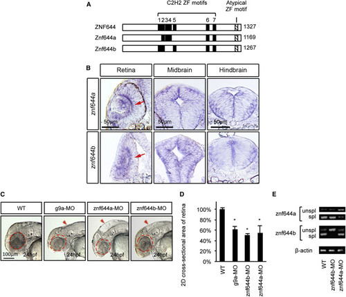
Zebrafish znf644 Paralogs Are Specifically Expressed In and Regulate the Development of Retinal and Midbrain Progenitor Cells (A) Conservation of C2H2-like ZF motifs between human and zebrafish ZNF644 genes. (B) Representative lateral views, retinal cross-sections, and midbrain cross-sections of whole-mount in situ hybridization (WISH) assays monitoring znf644a or znf644b expression at 24 hpf (n > 15 in each group). Arrows denote the inner part of the central retinal epithelium. (C) Lateral views of g9a (20/32), znf644a (10/18), or znf644b (14/21) morphant embryos compared with WT (21/21). Dashed circles denote 2D area of the WT retina. Arrowheads denote the dorsal hindbrain region. (D) Quantitation (mean ± SD) of retinal 2D cross-sectional area (¼m2) of znf644a, znf644b, or g9a morphants relative to WT embryos. Error bars represent SD. *p < 0.005, Student′s t test. (E) RT-PCR assays monitoring unspliced (unspl) and spliced (spl) znf644a or znf644b transcripts at 24 hpf. |
| Genes: | |
|---|---|
| Fish: | |
| Anatomical Terms: | |
| Stage: | Prim-5 |
| Fish: | |
|---|---|
| Knockdown Reagents: | |
| Observed In: | |
| Stage: | Prim-5 |