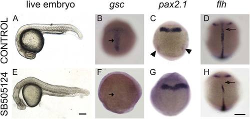
Exposure to Nodal inhibitor SB505124 at 3.8 hpf causes loss of mesendodermal and mesodermal derivatives. (A–D) Control WT embryos exposed to DMSO and (E–H) WT embryos exposed to 50 µM SB505124. (A) WT embryo at 1 dpf has a normal smooth and round head, a straight body axis, and complete somite formation, while (E) inhibitor-treated embryo displays cyclopia and abnormal morphology. (B) Normal gsc expression in the prechordal plate of a 9 hpf embryo (90% epiboly) displays a T-shaped expression domain (arrow) that (F) is absent in an inhibitor-treated embryo. (C) Normal pax2.1 expression is present along the midbrain–hindbrain border and in the posterior region in the pronephric mesoderm (arrowheads) in 10 hpf (tailbud stage) embryos. (G) In Nodal-deficient embryos, pax2.1 mRNA is present in the midbrain–hindbrain border (arrowheads) but absent in the pronephric mesoderm. (D) At 12 hpf (6 somite stage), flh is expressed in the developing notochord (arrow). (H) SB505124 treated embryos have partial reduction of notochord tissue. Scale bar: 250 µm.
|
