
Fig. 6
- ID
- ZDB-FIG-160225-8
- Publication
- Elkouby et al., 2016 - Oocyte Polarization Is Coupled to the Chromosomal Bouquet, a Conserved Polarized Nuclear Configuration in Meiosis
- Other Figures
- All Figure Page
- Back to All Figure Page
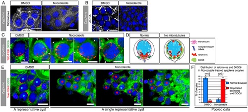
Nuclear telomere clustering and cytoplasmic Bb precursor localization are mechanistically coordinated by the bouquet microtubules.(A) Bouquet microtubules (Tg(βAct:emtb-3GFP), white) are intact in DMSO-treated ovaries (100%; n = 4 ovaries), but depolymerized in nocodazole treated ovaries (100%; n = 5 ovaries). A representative zygotene nest is shown (costained with DAPI, blue). Scale bar: 10 µm. (B) Acetylated tubulin cables (white) are intact in DMSO ovaries (100%; n = 3 ovaries), but are lost in nocodazole treated ovaries (100%; n = 6 ovaries; except for rare cases (3 nests) where cables were still intact, all other nest cables were disrupted). A representative zygotene nest is shown (costained with DAPI, blue). Scale bar: 10 µm. (C) Microtubules are simultaneously required for telomere clustering and DiOC6 localization at zygotene bouquet. DMSO zygotene oocytes (left panel) show the typical bouquet telomere cluster (Telo-FISH, red, red arrowheads) with the apposing localized DiOC6 (green, white arrowhead), similar to WT control (compare with Fig 4A). In contrast, nocodazole treatments (four right panels) resulted in simultaneous radial expansion of both telomeres and DiOC6. Four representative oocytes are shown. Note the partial to complete radial expansion of telomeres (red arrowheads) and the concomitant expansion and ectopic enrichments of DiOC6 (white arrowheads). Scale bar: 5 µm. (D) Schematics of the effects of the loss of microtubules on telomeres and DiOC6 distributions during zygotene bouquet. (E) Quantification of the results shown in (C). Zygotene oocytes were scored blind and mid-zygotene oocytes (11.5–13 µm) were selected for analysis. These were examined for their telomere clustering and DiOC6 patterns. A representative DMSO control nest is shown (left), where zygotene oocytes show the typical telomere clustering and apposing DiOC6 localization (nuclei labeled with blue dots). Partial projections of three different planes of a representative nocodazole-treated nest are shown (three right panels sequentially). In this nest, 10 oocytes showed radially-expanded telomeres as well as radial expansion, dispersion, or ectopic enrichments of DiOC6 (nuclei labeled with red dots), and one oocyte was normal (blue dot). Scale bar: 10 µm. (F) Pooled statistics of the analyzed nests as in (E). 93% (n = 45) of DMSO midzygotene oocytes were normal, while only 11.5% of midzygotene oocytes in nocodazole-treated ovaries were normal and 88.5% (n = 71) showed the effects described in (C, E). Bars are standard deviation (SD) of two independent experiments. Data in S1 Data. |