
Fig. S3
- ID
- ZDB-FIG-160225-11
- Publication
- Elkouby et al., 2016 - Oocyte Polarization Is Coupled to the Chromosomal Bouquet, a Conserved Polarized Nuclear Configuration in Meiosis
- Other Figures
- All Figure Page
- Back to All Figure Page
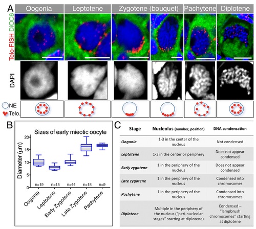
Precise criteria for staging early meiotic zebrafish oocytes.(A) Top: Images of the entire oocytes in Fig 2A, also showing DiOC6, which labels the cytoplasm and makes evident the size of the oocyte. Bottom: Nuclear zoom-in views of the same oocytes. DAPI (greyscale) show chromosome morphology. Range of sizes for specific stages is plotted in (B). Oocytes from 3–4 ovaries per stage were measured. Data in S1 Data. (C) Additional nuclear morphological criteria for each stage, including nucleolus number and positions, as well as DNA condensation, state characteristic of these early stages. These criteria expand upon the previously described characteristics of early oocytes in the zebrafish [38–40]. |