
Fig. 5
- ID
- ZDB-FIG-160225-7
- Publication
- Elkouby et al., 2016 - Oocyte Polarization Is Coupled to the Chromosomal Bouquet, a Conserved Polarized Nuclear Configuration in Meiosis
- Other Figures
- All Figure Page
- Back to All Figure Page
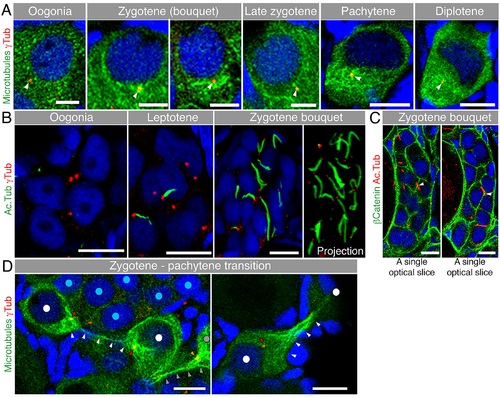
Microtubules in early meiosis.(A) Microtubule (Tg(βAct:emtb-3GFP), immunostained for GFP, green) organization during early meiotic oocytes costained with γTub (red) and DAPI (blue) (n = 6 ovaries). Scale bar: oogonia, zygotene = 5 µm; pachytene, diplotene = 10 µm. (B) Acetylated microtubule (Ac.Tub) cables in the nest. Ac.Tub cables (green) associate with centrosomes (red) of leptotene (center) and zygotene (right) oocytes (costained with DAPI), but are not present in oogonia (left) (n = 4 ovaries). Scale bar: 10µm. Right panel shows a full projection of Ac.Tub cables and centrosomes in the zygotene nest shown (see also S8 Video). (C) Some Ac.Tub cables (red) appeared to extend across the cell membranes (β-Catenin; green) of neighboring oocytes in the nest (costained with DAPI, blue; n = 12 ovaries). Arrowheads indicate crossing sites. Scale bar: 10 µm. Two single optical slices of the same nest are shown, see also S9 Video. (D) Zygotene to pachytene transitioning oocytes (DAPI, blue) show thin microtubule connections (Tg(βact:emtb-3GFP), green; white arrowheads) from the dense microtubule network around the centrosome (red; red arrowheads) of one cell to that of the other (n = 6 ovaries). The nuclei of the connected oocytes are labeled with white dots. Grey dot, grey and orange arrowheads indicate another oocyte with its centrosome and microtubule connection to an oocyte outside the image plane. Such oocytes were always observed in the periphery of a zygotene nest (nuclei labeled with blue dots, left panel; outside the image plane of right panel). Scale bar: 10 µm. Arrowheads indicate the centrosomes. |