
Fig. S2
- ID
- ZDB-FIG-160225-10
- Publication
- Elkouby et al., 2016 - Oocyte Polarization Is Coupled to the Chromosomal Bouquet, a Conserved Polarized Nuclear Configuration in Meiosis
- Other Figures
- All Figure Page
- Back to All Figure Page
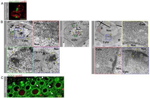
The nuclear cleft is a feature of the early oocyte.(A) The nuclear cleft, as visualized with LamB1, is enriched with the Bb precursor dazl RNA. Diplotene onset (top), pachytene (bottom). S2 Video shows the full stack of images of these oocytes, and S3 Video shows their 3-D view. (B) TEM images of representative ~20 µm (µ), ~20–25 µm and ~30 µm oocytes showing the typical nuclear cleft for these stages. The colors of framed regions in lower magnification images match the frame colors of the corresponding higher magnification images. Blue and green frames are higher magnification views of in-cleft cytoplasm. Red and yellow frames are higher magnification views of noncleft cytoplasm. The NE (yellow arrowheads) is concave forming the cleft, which is enriched with mitochondria (yellow arrows are examples of mitochondria) and electron-dense material presumably detecting mRNPs (*). Non-cleft cytoplasm contains presumptive mRNPs but is not enriched with mitochondria, and its adjacent NE is not concave. Cyto, cytoplasm; Nuc, nucleus. Note the nuclear peninsulas in the more pronounced cleft of the ~20 µm oocyte. In the ~20–25 µm oocyte, the cleft appears to be perpendicular to the image plane, with a view into the cleft. Note the cytoplasm (red *) engulfed by the nuclear protrusions and the presumptive mRNP clusters (green arrowheads) all around the NE inside the cleft. On the confocal microscope, as shown here in TEM, the nuclear cleft morphology of the 30 µm oocyte is consistently milder. Scale bars are indicated. (C) LamA/C is specifically detected in postcleft oocytes where the nucleus resumes a spherical shape (red arrowheads). Cleft stages are marked with white arrowheads. DiOC6 detects the concave NE and the cleft-enriched mitochondria. S4 Video shows the complete confocal stack. LamB1 is expressed in cleft (panel A; Fig 1C) and postcleft stages. |