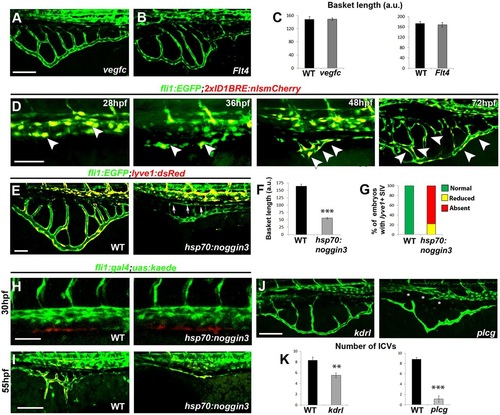
Molecular mechanisms controlling formation of the subintestinal plexus. (A-C) Subintestinal vessels form normally in vegfc (A,C) and flt4 (B,C) mutants. nvegfc=19; nFlt4=5. (D) Activation of BMP signaling is detected in the PCV (28hpf, arrowheads) and SIV (36-72hpf, arrowheads) of Tg(2xID1BRE:nlsmCherry) embryos. (E-G) Heat shock induction of noggin3 expression in Tg(fli1:EGFP; lyve1:dsRed; hsp70l:noggin3) embryos inhibits ventral migration of the plexus, as manifested by reduced basket length when compared with Tg(fli1:EGFP;lyve1:dsRed) (WT) embryos (E,F). The SIA forms normally (E, arrows). Cells in the SIV of noggin3-overexpressing embryos fail to upregulate the lymph-venous marker lyve1 (E,G). (H,I) The vPCV of Tg(fli1:gal4;uas:kaede) (WT) and Tg(fli1:gal4;uas:kaede;hsp70l:noggin3) double transgenic embryos was photoconverted at 30hpf (H). At 55hpf, red/yellow-labeled ECs were detected in the SIV of both WT and hsp70l:noggin3-overexpressing embryos (I). (J,K) Impaired Vegf signaling results in reduced number of ICVs in kdrl and plcg mutants (J,K). The SIA is absent in plcg mutants (J, asterisks).Yellow channel denotes colocalization of green and red fluorescence. Scale bars: 50µm. nvegfc=19; nFlt4=5; nnoggin3=11; nkdrl=13; nplcg=9. **P<0.01; ***P<0.001. Error bars represent s.e.m.
|
