
Fig. 2
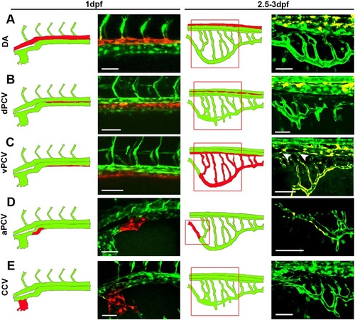
PCV cells give rise to all components of the subintestinal plexus. (A-E) Photoswitching of selected ECs was performed at 1dpf in Tg(fli1:gal4;uas:kaede) embryos, and vessels were scored for the presence of red-labeled ECs at 2.5-3dpf. Schematic illustrations of the corresponding confocal images are shown to the left. No photoconverted red cells were detected in the subintestinal plexus following photoswitching of the DA (A), dPCV (B) and CCV (E). Photoswitching of the vPCV (C) rendered red-labeled ECs in all components of the subintestinal plexus, including the SIA (arrowheads), whereas photoconverted ECs from the aPCV (D) were found exclusively in the rostral most part of the SIV. aPCV, anterior PCV; CCV, common cardinal vein; dPCV, dorsal PCV; vPCV, ventral PCV; for other abbreviations see legend to Fig. 1. Yellow channel denotes colocalization of green and red fluorescence. Scale bars: 50µm. nDA=24, ndPCV=10, nvPCV=16, naPCV=5, nCCV=11. |