
Fig. 3
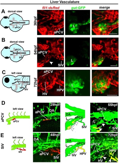
The left subintestinal vein and aPCV give rise to the liver vasculature. (A-C) Confocal images of Tg(gut:GFP;fli1:dsRed) double transgenic embryos at 36-72hpf highlighting the vasculature (red) and the liver (L, green). (A) The aPCV on the left side of the embryo is found adjacent to the liver at 36hpf. (B,C) ECs surround the liver by 54hpf (B, arrowhead) and form the HVs by 72hpf (C). The SIV drains into the liver via the HPV (C). (D,E) Photoswitching of ECs in the left branch of the aPCV (D) or the left SIV (E) was performed at 29hpf in Tg(fli1:gal4;uas:kaede) embryos. (D) Red-labeled ECs from the left aPCV contribute to the liver vasculature (55hpf, arrowheads). (E) Photoswitching of the left SIV at 48hpf rendered red-labeled ECs in the HVs and the HPV (E, 72hpf, arrowheads). aPCV, anterior PCV; HPV, hepatic portal vein; HV, hepatic vessels; L, liver; for other abbreviations see legend to Fig. 1. Yellow channel denotes colocalization of green and red fluorescence. Scale bars, 50µm. naPCV=4, nSIV=5. |