
Fig. 4
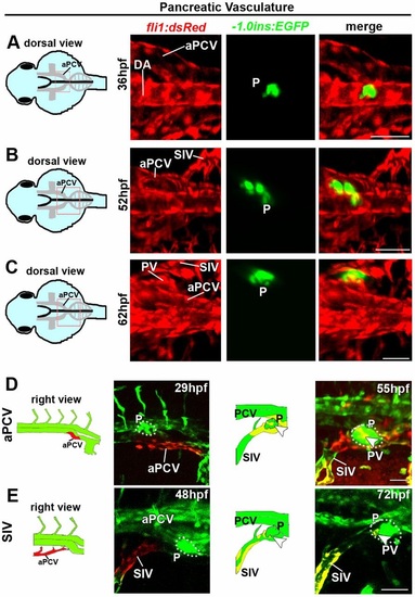
The right subintestinal vein and aPCV give rise to the pancreatic vasculature. (A-C) Confocal images of Tg(-1.0ins:EGFP;fli1:dsRed) embryos at 36-62hpf highlighting the vasculature and the endocrine pancreas (P, green). (A) insulin:EGFP+ cells are detected between the two branches of the aPCV at 36hpf. (B,C) Gradual shift of insulin:EGFP+ cells towards the right side of the midline. The most cranial ICV on the right side of the plexus (C) sends branches towards the nascent pancreas and forms pancreatic vessels (PV). (D,E) Photoswitching of ECs in the right branch of the aPCV at 29hpf (D) or in the right SIV at 48hpf (E) was performed in Tg(fli1:gal4;uas:kaede;-1.0ins:EGFP) embryos. (D) Red-labeled ECs from the right aPCV contribute to the pancreatic vessels (PV) (55hpf, arrowhead). (E) Photoswitching of the right SIV at 48hpf rendered red-labeled ECs in the pancreatic vessels (PV) (72hpf, arrowheads). aPCV, anterior PCV; P, pancreas; PV, pancreatic vessels; for other abbreviations see legend to Fig. 1. Yellow channel denotes colocalization of green and red fluorescence. Scale bars, 50µm. naPCV=8, nSIV=11. |