
FIGURE
Fig. S3
Fig. S3
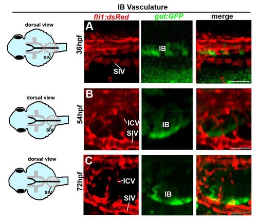
The intestinal bulb develops in close contact to the subintestinal vessels (A-C) Confocal images of Tg(gut:GFP;fli1:dsRed) embryos at 36-72 hpf showing morphogenesis and growth of the intestinal bulb (IB) and its vasculature arising from the ICVs and SIV. ICV, interconnecting vessel; IB, intestinal bulb; SIV, subintestinal vein. Scale bars: 50µm. |
Expression Data
Expression Detail
Antibody Labeling
Phenotype Data
Phenotype Detail
Acknowledgments
This image is the copyrighted work of the attributed author or publisher, and
ZFIN has permission only to display this image to its users.
Additional permissions should be obtained from the applicable author or publisher of the image.
Full text @ Development