
Fig. 5
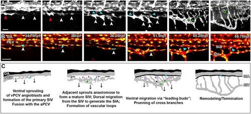
Formation of the subintestinal plexus involves different mechanisms of EC migration. (A) Snapshots from a time-lapse sequence of a Tg(fli1:dsRed) embryo, showing individual sprouts arising from the vPCV (32hpf, white arrowheads), which quickly anastomose and fuse with a caudal projection of the aPCV (red arrowhead) to generate the primary SIV. Sprouts arising along the primary SIV elongate ventrally and fuse to generate the mature SIV (35-41hpf, white arrowheads). Concomitantly, ECs from the SIV migrate dorsally to generate the SIA (39-41hpf, light-blue arrowheads). The mature SIV migrates ventrally through collective migration guided by leading buds (56hpf, white arrows), while fusion of angiogenic sprouts originating in the primary SIV generates vascular loops (56hpf, green asterisks). Retraction of the leading buds, along with pruning of the cross-branches forming the vascular loops, render the stereotypical basket shape (78hpf) that engulfs the intestinal bulb (IB). (B) Snapshots from a time-lapse sequence of a Tg(fli1:dsRed;fli1:nGFP) embryo showing the migration route taken by vPCV angioblasts. A single angioblast (light blue) initially residing in the vPCV (34.75hpf, white arrowhead) sprouts ventrally (36hpf, white arrowhead) and incorporates into the primary SIV (36.25-51.5hpf, white arrowhead). Later on, the same cell migrates dorsally (55.25hpf, light blue arrowhead), eventually becoming incorporated into the SIA (59.75hpf, light blue arrowhead). (C) Schematic illustration depicting the different steps involved in formation of the subintestinal plexus: ventral sprouting and ventral migration (black arrows); cells ‘lagging back’ followed by dorsal migration (purple arrows); vascular loops (green asterisks); and vessel pruning (black arrowhead). Scale bars: 50µm. |