
FIGURE
Fig. S13
Fig. S13
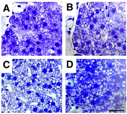
Liver structure is not altered in acute organophosphorus poisoning in zebrafish. Normal hepatic structures and normal hepatocytes are observed both in 8 days post-fertilization control (A), grade 1 (B), grade 2 (C) and grade 3 (D) zebrafish larvae. Hepatocyte nuclear size may not be homogeneous in the same fish and darker nuclei in D correspond to erythroblasts/erythrocytes. The presence of lipid droplets (white spherical structures) is a normal observation corresponding to yolk remnants, and their number may not always be homogeneous in fish from the same batch. Scale bar: 80 µm. |
Expression Data
Expression Detail
Antibody Labeling
Phenotype Data
Phenotype Detail
Acknowledgments
This image is the copyrighted work of the attributed author or publisher, and
ZFIN has permission only to display this image to its users.
Additional permissions should be obtained from the applicable author or publisher of the image.
Full text @ Sci. Rep.