|
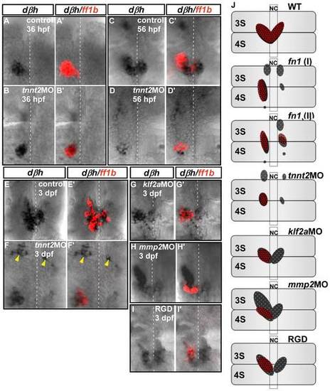
Interaction between interrenal steroidogenic and chromaffin cells in tnnt2a, klf2a, and mmp2 morphants and RGD-treated embryos during interrenal gland assembly. Double ISH assays showing colocalization of ff1b (red) and dbh (black) transcripts in uninjected control embryos and tnnt2a morphants at (A, A&prime& B, B′) 36 hpf (n = 3 and 6, respectively), (C, C&prime& D, D′) 56 hpf (n = 3 and 5, respectively), and (E, E&prime& F, F′) 3 dpf (n = 17 and 15, respectively), and in (G, G′) klf2a (n = 9) and (H, H′) mmp2 (n = 8) morphants and (I, I′) RGD-treated embryos (n = 18) at 3 dpf. Ventral flat mount views are shown for representative embryos in each group, oriented with anterior at the top. Yellow arrowheads indicate chromaffin cell clusters that failed to converge at the interrenal area in tnnt2a morphants. Broken white lines indicate position of the midline. (J) Schematic representation of various phenotypic defects associated with interrenal organ assembly. Panels show ventral views of wild-type, mutant, morphant, and drug-treated embryos at 3 dpf, oriented with anterior at the top. Phenotypes depicted for cloche (clo) and fn1 mutants are based on previous reports [9], [21]. Abbreviations: notochord (NC), the third somite (3S), the fourth somite (4S).
|