FIGURE

Fig. 8

ID
ZDB-FIG-150325-19
Publication
Mommaerts et al., 2014 - Smoc2 modulates embryonic myelopoiesis during zebrafish development
Other Figures
All Figure Page
Back to All Figure Page
Fig. 8

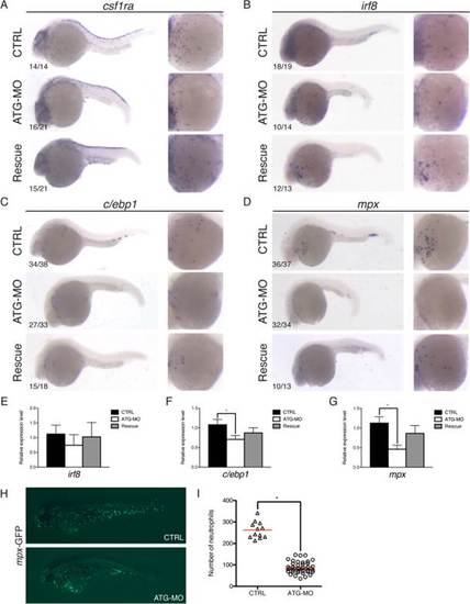
smoc2 morphants display defects in embryonic myelopoiesis at the onset of blood circulation. A–D: Lateral views of the embryos, anterior to the left, dorsal to the top, at the onset of circulation. A: WISH analysis or the macrophage markers csf1ra (A) and irf8 (B), and the neutrophil markers c/ebp1 (C) and mpx (D). E–G: qPCR analysis for irf8 (E), c/ebp1 (F) and mpx (G). Values plotted as mean ± SEM; n = 4; *P < 0.05. H,I: Visualization of the mpx-positive cells using the mpx reporter embryos (Tg(mpx:GFP)i114) at 3 dpf (H) and the quantification of the number of neutrophils in the ATG morphants (n = 42) as compared to the control embryos (n = 12; *P < 0.05) (I).

Expression Data
Genes:
Fish:
Knockdown Reagents:
Anatomical Terms:
Stage Range: Prim-5 to Protruding-mouth

Expression Detail
Antibody Labeling
Phenotype Data
Fish:
Knockdown Reagents:
Observed In:
Stage Range: Prim-5 to Protruding-mouth

Phenotype Detail
Acknowledgments
This image is the copyrighted work of the attributed author or publisher, and ZFIN has permission only to display this image to its users. Additional permissions should be obtained from the applicable author or publisher of the image. Full text @ Dev. Dyn.