
Fig. 1
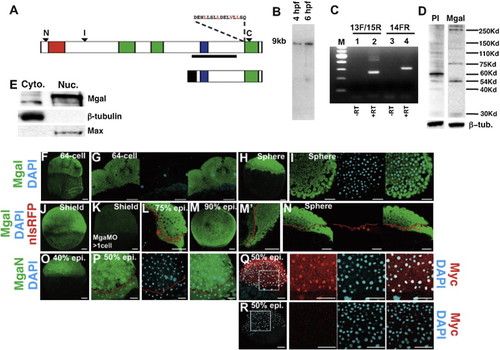
MGA Protein Is Maternally and Zygotically Expressed in All Cells (A) Diagram of full-length MGA protein (top) or a C-terminal MGA fragment consisting of amino acids 2,058–2,735 (bottom). Arrowheads indicate the location of the epitopes for anti-MGA antibodies. A conserved leucine rich nuclear export signal is shown. T-domain (residues 105–285), red; bHLHZip domain (residues 2,093–2,186), blue; Conserved Region 1 (CR1) (residues 938–1,166), CR2 (residues 1,260–1,370), CR3 (residues 2,540–2,715), green; Myc-epitope, black. (B) Northern blot of 4 hpf and 6 hpf embryos for mga transcripts, using the probe indicated by the line in (A). (C) RT-PCR of 8 hpf embryo extracts using primers anchored on either side of exon 14 (13F/15R), or internal to exon 14 (14FR). The lower (260 bp) band in lane 2 lacks exon 14; the upper bands (450 bp and 500 bp) contain exon 14. The 300 bp product in lane confirms the presence of exon 14. (D) Western blot of 8 hpf embryo extracts showing specific bands at 290 kDa and 250 kDa. (E) Western blot of cytoplasmic and nuclear extracts from 8 hpf embryos. (F–P) Confocal images of MGA protein distribution. Embryos were counterstained with DAPI or nls-RFP as indicated, and the boundary between the YSL and blastoderm is marked by a red line in (L), (M2), and (N). (Q and R) Confocal images showing distribution of anti-Myc epitope in embryos injected with C-terminal MGA fragment shown in (A) or uninjected embryos; counterstained with DAPI. Dorsal is to the right in (H), (I), (L), and (L2). At other stages there are no morphological landmarks to determine the orientation. Scale bar represents 50 μm. See also Figure S3. |
| Gene: | |
|---|---|
| Antibodies: | |
| Fish: | |
| Knockdown Reagents: | |
| Anatomical Terms: | |
| Stage Range: | 64-cell to 90%-epiboly |
Reprinted from Developmental Cell, 28(3), Sun, Y., Tseng, W.C., Fan, X., Ball, R., and Dougan, S.T., Extraembryonic signals under the control of MGA, Max, and Smad4 are required for dorsoventral patterning, 322-334, Copyright (2014) with permission from Elsevier. Full text @ Dev. Cell