FIGURE

Fig. 2

ID
ZDB-FIG-140305-57
Publication
Haddon et al., 1998 - Multiple delta genes and lateral inhibition in zebrafish primary neurogenesis
Other Figures
All Figure Page
Back to All Figure Page
Fig. 2

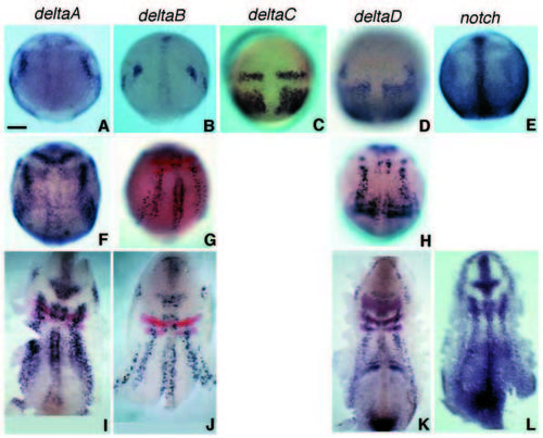
Early expression patterns of all four zebrafish delta genes and of notch seen by in situ hybridisation. Dorsal views, anterior to the top. (A-E) At 90% epiboly (9 hpf); (F-L) at bud to 1-somite stage (10-10.5 hpf). The middle row (FH) shows intact embryos, the bottom row (I-L), dissected flat mounts. Red stain in G and in I-K shows expression of krox20 and, in K, of paxb. Tissue sections (not shown) confirm that deltaA and deltaB are expressed in the epiblast, in prospective neural tissue, whereas the strong expression of deltaC and deltaD at these stages is in the prospective mesoderm. Scale bar: 100 μm.

Expression Data
Genes:
Fish:
Anatomical Terms:
Stage Range: 90%-epiboly to 1-4 somites

Expression Detail
Antibody Labeling
Phenotype Data

Phenotype Detail
Acknowledgments
This image is the copyrighted work of the attributed author or publisher, and ZFIN has permission only to display this image to its users. Additional permissions should be obtained from the applicable author or publisher of the image. Full text @ Development