Fig. 2
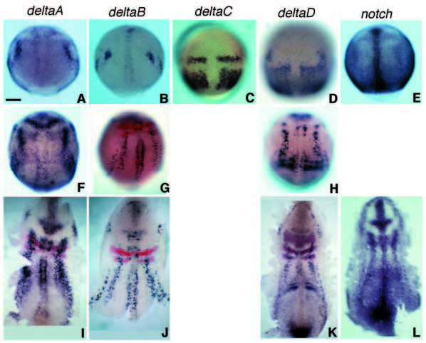
Fig. 2 Early expression patterns of all four zebrafish delta genes and of notch seen by in situ hybridisation. Dorsal views, anterior to the top. (A-E) At 90% epiboly (9 hpf); (F-L) at bud to 1-somite stage (10-10.5 hpf). The middle row (FH) shows intact embryos, the bottom row (I-L), dissected flat mounts. Red stain in G and in I-K shows expression of krox20 and, in K, of paxb. Tissue sections (not shown) confirm that deltaA and deltaB are expressed in the epiblast, in prospective neural tissue, whereas the strong expression of deltaC and deltaD at these stages is in the prospective mesoderm. Scale bar: 100 μm.

