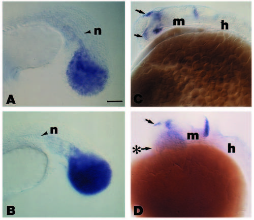
Expression pattern of wnt8 (A) and snail1 (B) in the tailbud of spadetail mutant embryos, and wnt8b in the rostral CNS of wild-type (C) and cyclopsb16 mutant (D) embryos. Anterior is to the left and dorsal is to the top in all photomicrographs. (A) For wnt8, the increase in the relative intensity of the signal compared with that in a wild-type embryo (Fig. 3F), can be attributed to the snail1-positive cells (Fig. 5B) that failed to converge properly during gastrulation. (C) In wild-type embryos at approximately 24 hours postfertilization, wnt8b is expressed in the epiphysis (top arrow), the optic stalk (lower arrow), the ventral floor of the forebrain, just caudal to the optic stalk signal, and at the midbrain and hindbrain junction. (D) In homozygous cyc-/- mutants, wnt8b expression is unaffected in the epiphysis (top arrow) and at the boundary behind midbrain and hindbrain. Note the loss of the signal in the optic stalk (arrow from asterisk) and in the ventral floor of the forebrain just caudal to it. Abbreviations: h, hindbrain; m, midbrain; n, notochord. Scale bar (A,B), 45 μm; (C,D), 55 μm.
|
