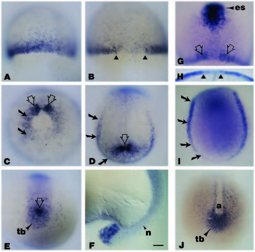
Whole-mount in situ hybridization analyses of the spatial localization of wnt8 during early gastrulation to midsegmentation. (A) Embryo at 50% epiboly illustrating the wnt8 signal in cells at the blastoderm margin; animal pole is to the top. (B) Dorsal view (animal pole to the top), of a whole-mount embryo at approximately 75-80% epiboly. Note the absence of the wnt8 signal in the dorsal midline (triangles). (C) Vegetal pole view of a whole-mount embryo at late gastrulation (90-95% epiboly); dorsal is to the top. The wnt8 signal, no longer evident on the ventral side and downregulated on the lateral sides (black arrows), becomes enriched on either side of the dorsal axial mesoderm (open arrows). (D) During early segmentation (approximately 10-11 hours), the wnt8 signal, seen in this dorsal view of a whole-mount embryo where anterior is to the top, is confined to the lateral and caudal margin of the embryonic shield (black arrows), and in the area surrounding the presumptive tailbud (open arrow). (E) Vegetal pole view illustrating the wnt8 pattern of expression at the posterior end of an embryo at the 1- to 2-somite stage. Note that the signal is now restricted to the tailbud and the cells immediately adjacent to it (open arrow). (F) At 16-18 hours, the wnt8 signal is obvious at the tip of the tail. (G) Dorsal view of a whole-mount embryo at approximately 80% epiboly, illustrating the expression pattern of wnt8 and gsc. The expression of gsc, in cells of the anterior embryonic shield (es), delineates the wnt8- negative area corresponding to the axial hypoblast (open arrows). (H) Higher magnification view from the animal pole clearly demonstrates that by late gastrulation, wnt8 is no longer expressed in cells in the dorsal midline (triangles). (I) Dorsal view with anterior to the top of a whole-mount embryo stained for pax2 expression. Note that the pax2 signals in the lateral and caudal margins of the shield (arrows), are similar to those in a comparable stage embryo stained for wnt8 (Fig. 3D). (J) The expression pattern of snail1 in a whole-mount embryo at a stage comparable to the one in Fig. 2E. Although wnt8 (Fig. 3E) and snail1 (Fig. 3J) are expressed in the tailbud, only snail1 is expressed in the more anterior regions flanking the midline axis. Abbreviations: a, axial mesoderm; n, notochord; tb, tailbud. Scale bar (A-E, G-J), 55 μm; (F), 45 μm.
|
