Fig. 6
- ID
- ZDB-FIG-140227-25
- Publication
- Kelly et al., 1995 - Zebrafish wnt8 and wnt8b share a common activity but are involved in distinct developmental pathways
- Other Figures
- All Figure Page
- Back to All Figure Page
|
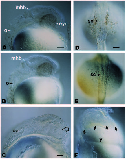
Morphological comparisons between uninjected, β-galactosidase-RNA-injected and wnt8-RNA-injected zebrafish embryos at approximately 30 hours postfertilization. Embryos in (A-C) are oriented anterior to the top and dorsal to the left. (A) The prominent eye, otocyst and midbrain-hindbrain boundary are evident in this lateral view of an uninjected embryo. (B) Note the apparent normal morphology of these structures in a lateral view of an embryo having been injected with approximately 1 ng of b-galactosidase RNA. (C) In embryos injected with approximately 0.1 ng of wnt8 RNA, the region where the eye should have formed (arrow) and the regions flanking the midbrain-hindbrain boundary are noticeably affected, but the otocyst appears to have developed normally. (D) Dorsal view of an uninjected embryo at approximately 30 hours, illustrating the spinal cord oriented in an anterior (top) to posterior (bottom) direction. (E) Dorsal view of an embryo having been injected with approximately 0.2 ng of wnt8 RNA, illustrating that a single axis forms even though there were defects apparent in the mid- and forebrain similar to those seen in C, but out of the plane of focus. The spinal cord in these wnt8-injected embryos is morphologically similar to that seen in the uninjected control (D). (F) Lateral view of an embryo having been injected with approximately 0.2 ng of wnt8 RNA and examined at 18 hours of development. The majority of this embryo has developed as an amorphous mass above the yolk (arrows). Despite the few anatomically visible landmarks, and although it is out of the plane of focus, there is a morphologically recognizable notochord exhibiting the characteristic vacuolated arrangement (see Westerfield, 1989). Abbreviations: mhb, midbrain-hindbrain boundary; o, otocyst; sc, spinal cord; y, yolk. Scale bar (A, B), 55 μm; (C), 40 μm; (D-F), 65 μm. |

