
Fig. S1
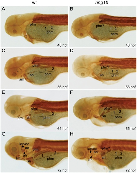
Cranial musculature development is severely impaired in ring1b mutants. Lateral views of embryonic musculature in WT and ring1b mutants (A–H). The posterior hypaxial muscle (phm), had delaminated from the somites in both WT and ring1b mutants at 48 hpf (A, B). At 56 hpf, the sternohyoideus (sh) is formed in both WT and ring1b mutants and the pectoral fin muscle is prominently visible (C, D). During later development, the sh and phm elongated and attached to the cleithrum, a bone of the fin girdle in WT embryos, (E, G). Elongation of these muscles was completely abrogated in ring1b mutants (F, H). Cranial musculature is almost completely absent in ring1b mutants (H). Abbreviations: am: anterior mandibularis; ah: adductor hyomandibulae; ao: adductor opercule; do: dilator operculi; hh: hyohyoideus; ih: interhyoideus; ima: intermandibularis anterioris; imp: intermandibularis posterioris, lap: levator arcus palatine; phm: posterior hypaxial muscle; pfm: pectoral fin muscle; sh: sternohyoideus. |