
Fig. 8
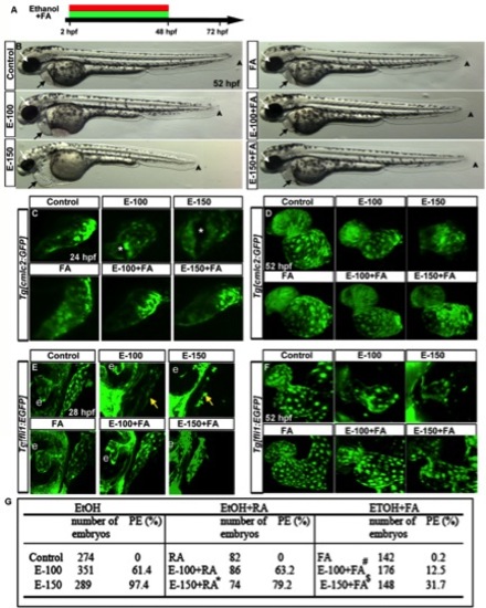
FA supplementation during chronic ethanol exposure (2–48hpf) rescues ethanol-induced cardiac defects. A: Schematic diagram showing the timing of ethanol and FA exposure. B: Live images showed heart edema, small eye, and short body length in the ethanol-exposed embryos and normal morphology in FA cosupplemented (2–48hpf) embryos. Black arrow, heart; white arrow, eye; arrowhead, posterior end of the embryo. C, D: Confocal images of Tg[cmlc2:GFP] embryos showed linear heart tube (24 hpf) and normal chambers (52 hpf) in the control, FA-treated, and ethanol+FA-cotreated embryos. Ethanol-exposed embryos exhibited abnormal heart tube with defective myocardial fusion (asterisk, 24 hpf) and misshapen hearts (52 hpf). E, F: Confocal images of Tg[fli1:EGFP] at 28 and 52 hpf showed misshapen endocardium (yellow arrows; e, eye) in the ethanol-treated; normal endocardium in control, FA-treated, and E-100+FA-cotreated; and near normal endocardium in E-150+FA-cotreated embryos. G: Scores showing the effect of continuous supplement of RA or FA with ethanol on pericardial edema (PE). Results shown were calculated from three individual experiments. (Mantel-Haenszel chi-square tests: control vs. EtOH-100 or EtOH-150, P < 0.0001; control vs. RA, P = 1.00; EtOH-150 vs. EtOH-150+RA, P < 0.0001 (*); control vs. FA, P = 0.01; EtOH-100 vs. EtOH-100+FA, P < 0.0001 (#); EtOH-150 vs. EtOH-150+FA ml, P < 0.0001 ($)). |