
Fig. 2
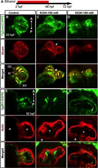
Chronic ethanol exposure during cardiogenesis produces defective chambers and endocardial cushion. A: Schematic diagram showing the developmental timing (2–48 hpf) of ethanol exposure. B–D: Ethanol-treated embryos do not have stereotypical regionally confined cardiomyocyte shapes. Extended focus images of confocal sections from control Tg[cmlc2:GFP] embryo (B) costained with alcam antibody showed large, elongated cardiomyocytes in the outer curvature (OC) and AV region, and small rounded cardiomyocyte in the inner curvature (IC) of the ventriclular surface in control embryos; the orientation of the cells was aligned with each other. C: Cardiomyocytes were disorganized, showing variable sizes and shapes in ethanol treated embryos. D: Ethanol treatment occasionally caused cardia bifida, which display small cardiomyocytes. Representative cell shapes are highlighted in yellow; white arrow, defective ventricular wall; white arrowhead, abnormal thin cell. E–G: Severely defective endocardial cushion formation was seen after ethanol treatment. E: Confocal sections of the control 52-hpf Tg[fli1:EGFP] embryo heart co-labeled with Texas red-phalloidin (F-actin) showed clusters of GFP- and F-actin-positive cells (white dotted line) at the AV boundary, which were reduced (F) or absent (G) in ethanol treated embryos. A, anterior; P, posterior; a, atrium; v, ventricle. |