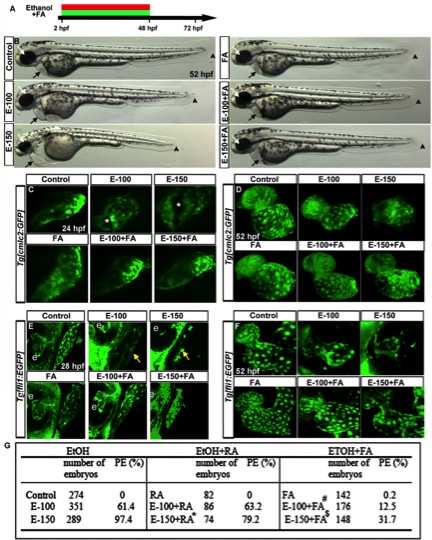
Fig. 8 FA supplementation during chronic ethanol exposure (2–48hpf) rescues ethanol-induced cardiac defects. A: Schematic diagram showing the timing of ethanol and FA exposure. B: Live images showed heart edema, small eye, and short body length in the ethanol-exposed embryos and normal morphology in FA cosupplemented (2–48hpf) embryos. Black arrow, heart; white arrow, eye; arrowhead, posterior end of the embryo. C, D: Confocal images of Tg[cmlc2:GFP] embryos showed linear heart tube (24 hpf) and normal chambers (52 hpf) in the control, FA-treated, and ethanol+FA-cotreated embryos. Ethanol-exposed embryos exhibited abnormal heart tube with defective myocardial fusion (asterisk, 24 hpf) and misshapen hearts (52 hpf). E, F: Confocal images of Tg[fli1:EGFP] at 28 and 52 hpf showed misshapen endocardium (yellow arrows; e, eye) in the ethanol-treated; normal endocardium in control, FA-treated, and E-100+FA-cotreated; and near normal endocardium in E-150+FA-cotreated embryos. G: Scores showing the effect of continuous supplement of RA or FA with ethanol on pericardial edema (PE). Results shown were calculated from three individual experiments. (Mantel-Haenszel chi-square tests: control vs. EtOH-100 or EtOH-150, P < 0.0001; control vs. RA, P = 1.00; EtOH-150 vs. EtOH-150+RA, P < 0.0001 (*); control vs. FA, P = 0.01; EtOH-100 vs. EtOH-100+FA, P < 0.0001 (#); EtOH-150 vs. EtOH-150+FA ml, P < 0.0001 ($)).
Image
Figure Caption
Acknowledgments
This image is the copyrighted work of the attributed author or publisher, and
ZFIN has permission only to display this image to its users.
Additional permissions should be obtained from the applicable author or publisher of the image.
Full text @ Dev. Dyn.

