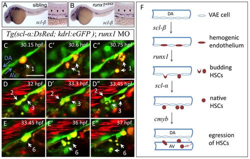
Scl-β, Runx1 and Scl-α regulate sequential steps of AGM HSC development. (A,B) Hemogenic endothelium marked by scl-β expression in 28 hpf siblings (heterozygotes and wild type) and runx1w84x mutant zebrafish embryos. Arrowheads indicate expression of scl-β in the AGM region. (C-E′) Time-lapse confocal imaging of a live Tg(scl-α:DsRed; kdrl:eGFP) embryo injected with runx1 MO from 32 to 39 hpf. White arrows indicate four scl-α:DsRed+ VAE cells that initiate budding but then fragment. Also see supplementary material Movie 5. Similar results were obtained in all three of the runx1 morphants examined and a total of 13 such events were observed (n=3/3). Scale bars: 20 μm. (F) A model of the molecular regulation at consecutive steps during the development of HSCs in the AGM. The establishment of hemogenic endothelium relies on scl-β. runx1 is crucial for their subsequent transformation into HSCs via EHT. The maintenance of nascent HSCs in the AGM requires scl-α, and cmyb is essential to regulate the egression of these HSCs from the AGM.
|
