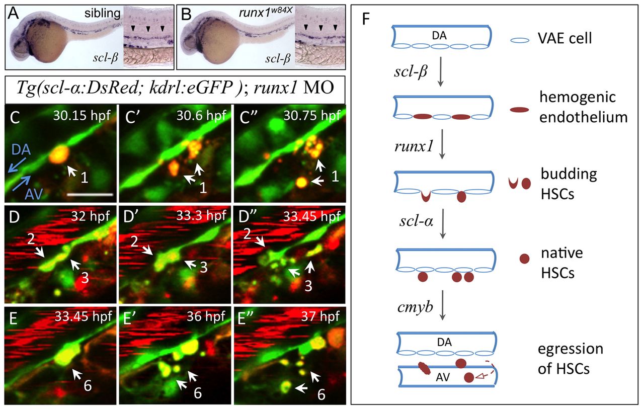
Fig. 7 Scl-β, Runx1 and Scl-α regulate sequential steps of AGM HSC development. (A,B) Hemogenic endothelium marked by scl-β expression in 28 hpf siblings (heterozygotes and wild type) and runx1w84x mutant zebrafish embryos. Arrowheads indicate expression of scl-β in the AGM region. (C-E′) Time-lapse confocal imaging of a live Tg(scl-α:DsRed; kdrl:eGFP) embryo injected with runx1 MO from 32 to 39 hpf. White arrows indicate four scl-α:DsRed+ VAE cells that initiate budding but then fragment. Also see supplementary material Movie 5. Similar results were obtained in all three of the runx1 morphants examined and a total of 13 such events were observed (n=3/3). Scale bars: 20 μm. (F) A model of the molecular regulation at consecutive steps during the development of HSCs in the AGM. The establishment of hemogenic endothelium relies on scl-β. runx1 is crucial for their subsequent transformation into HSCs via EHT. The maintenance of nascent HSCs in the AGM requires scl-α, and cmyb is essential to regulate the egression of these HSCs from the AGM.
Image
Figure Caption
Figure Data
Acknowledgments
This image is the copyrighted work of the attributed author or publisher, and
ZFIN has permission only to display this image to its users.
Additional permissions should be obtained from the applicable author or publisher of the image.
Full text @ Development

