|
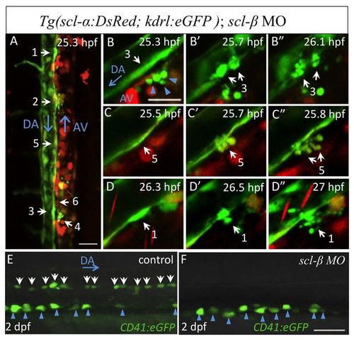
scl-β is crucial for hemogenic endothelium specification. (A-D′) Time-lapse confocal imaging of a live Tg(scl-α:DsRed; kdrl:eGFP) scl-β zebrafish morphant from 25 to 36 hpf. White arrows and numbers indicate the endothelial cells that will undergo fragmentation during the following 6 hours. In B-D′, three kdrl:eGFP+ elongated endothelial cells burst into fragments without initiation of budding. Also see supplementary material Movie 3. Blue arrowheads in B indicate some cell debris that already existed before recording. Similar results were obtained in all three scl-β morphants observed (n=3/3). (E,F) CD41:eGFP+ HSCs in the AGM region of 2 dpf live control embryos and scl-β morphants. On average, fewer than one CD41:eGFP+ HSC could be detected in the AGM of scl-β morphants (n=17/19), compared with an average of 13 CD41:eGFP+ HSCs (white arrows) in the same region of control embryos. Blue arrowheads identify pronephric duct cells. Scale bars: 30 μm in A; 20 μm in B-F.
|