
FIGURE
Fig. S1
Fig. S1
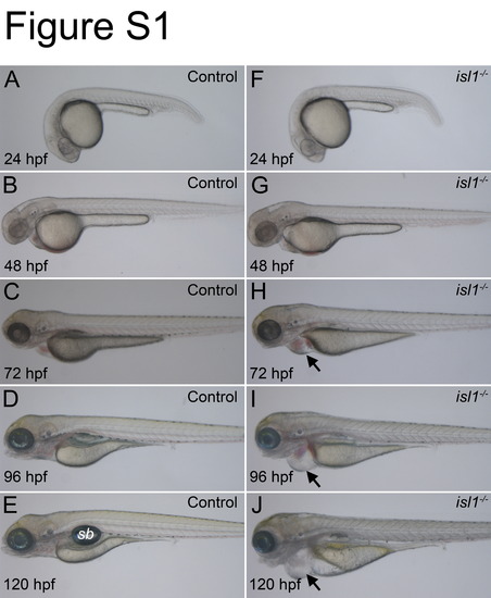
Phenotype of isl1 mutant zebrafish compared to wildtype. Images of live control embryos (A–E) and isl1 mutant embryos (F–J) taken between 24–120 hpf. (F–J) Mutant embryos appear normal during the first two days after fertilization but show heart edema at 72 hpf (black arrow head in H, I and J). At later stages, the isl1 mutant fish show various defects including smaller eyes, malformed somites and a non-inflated swim bladder (sb). |
Expression Data
Expression Detail
Antibody Labeling
Phenotype Data
Phenotype Detail
Acknowledgments
This image is the copyrighted work of the attributed author or publisher, and
ZFIN has permission only to display this image to its users.
Additional permissions should be obtained from the applicable author or publisher of the image.
Reprinted from Developmental Biology, 378(1), Wilfinger, A., Arkhipova, V., and Meyer, D., Cell type and tissue specific function of islet genes in zebrafish pancreas development, 25-37, Copyright (2013) with permission from Elsevier. Full text @ Dev. Biol.