
Fig. 8
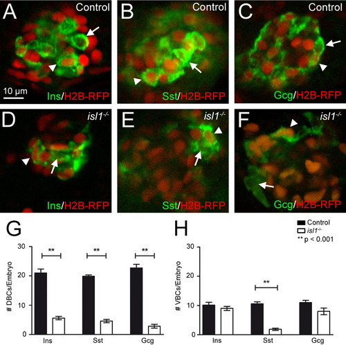
Endocrine hormone expression in first and second wave endocrine cells in control and isl1 mutant embryos. Single plane views from confocal images of the primary islets from control (A–C) and isl1 mutants (D–E). Embyros were injected with H2B-RFP (in red) encoding mRNA at the one cell stage and were immunostained (in green) for Ins (A, D), Sst (B, E) and Gcg (C, F) at 96 hpf. Cells that left cell-cycle early in development show a red nuclear label due to the injecetion of H2B-RFP encoding mRNA at the one cell stage. Arrowheads indicate examples of hormone+/RFP+ cells (refered to as DBCs), while arrows indicates examples of hormone+/RFP- cells (referred to as VBCs). (G, H) Quantification of hormone+ DBCs (G) and VBCs (H) in control (black bars) and isl1 mutant embryos (white bars). Note that isl1 mutants show significantly reduced numbers for all DBCs, while only the number of Sst expressing VBCs cells is significantly reduced. Bars show mean+SEM. |
Reprinted from Developmental Biology, 378(1), Wilfinger, A., Arkhipova, V., and Meyer, D., Cell type and tissue specific function of islet genes in zebrafish pancreas development, 25-37, Copyright (2013) with permission from Elsevier. Full text @ Dev. Biol.