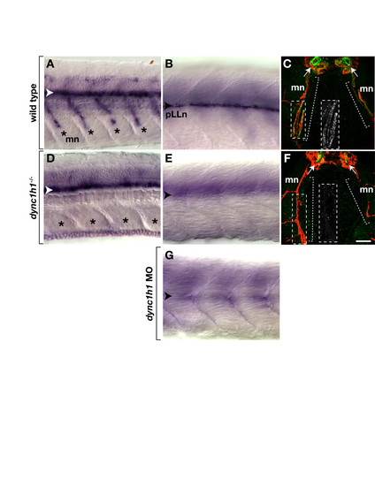
dync1h1 -deficient larvae express mbp RNA and protein in the central nervous system (CNS) but not the peripheral nervous system (PNS). (A, B) mbp RNA expression in the ventral spinal cord (white arrowhead), along motor nerves (mn, asterisks) and at the posterior lateral line nerve (pLLn, black arrowhead) of wild-type larvae. (C) Immunohistochemistry reveals Mbp (green) associated with motor nerves marked by Acetylated Tubulin staining (red, brackets). Arrows indicate prominent Mbp in the ventral spinal cord. The inset shows Mbp expression, alone, of the portion of the motor nerve, indicated by the dashed box. (D, E) mbp RNA expression in the spinal cord but not at the motor nerves or the pLLn of dync1h1 mutant larvae. (F) Mbp is absent from motor nerves and reduced in the ventral spinal cord of a mutant larva. The inset shows Mbp labeling, alone, of the portion of the motor nerve indicated by the dashed box. (G) pLLn of a wild-type larvae injected with dync1h1 antisense MO lacks mbp RNA expression. Panels A, B, D, E and G show whole 4.25 days post fertilization (dpf) larvae at the level of the mid-trunk with anterior to the left and dorsal to the top. Panels C and F show transverse sections through the level of the trunk with dorsal up. Scale bars equal 20 μm (A, B, D, E, G), 40 μm (C, F) and 50 μm for the insets.
|
