|
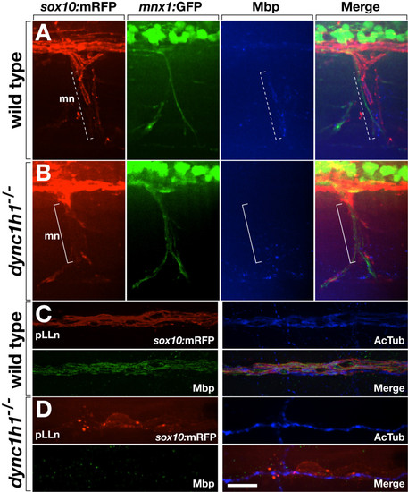
Loss of dync1h1 function disrupts axon wrapping and Mbp expression by Schwann cells. (A, B) Wild-type and dync1h1 mutant larvae expressing sox10:mRFP (red) and mnx1:GFP (green) and labeled by anti-Mbp antibody (blue). Tube-like Mbp+ Schwann cells are evident at some axons of the motor nerve (mn) in wild type (dashed bracket) whereas Mbp– Schwann cells appear more loosely associated with axons in the mutant (solid bracket). (C, D) Wild-type and dync1h1 mutant larvae expressing sox10:mRFP (red) and labeled by anti-Mbp (green) and anti-acetylated tubulin (AcTub, blue) antibodies. Mbp+ Schwann cells ensheath pLLn axons in wild-type. By contrast, RFP+ Schwann cells associated with the pLLn in dync1h1/ mutants do not express Mbp and appear loosely associated with axons. All panels show lateral views of 6 dpf wild-type and dync1h1/ larvae with anterior to the left and dorsal to the top. Scale bars equal 20 μm (A, B) and 10 μm (C, D).
|