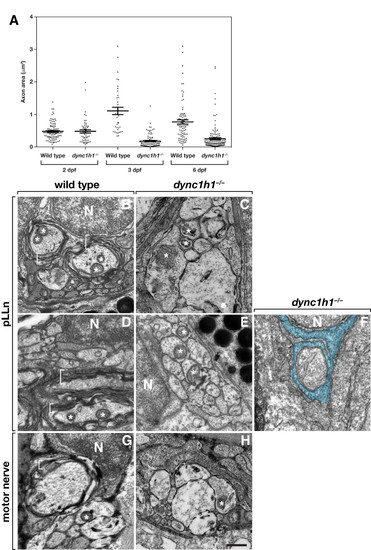
Dync1h1 is required for the formation of myelinating Schwann cells. (A) Scatter plot analysis of axon area. Each point represents one axon. Horizontal bars indicate mean axon area with SD for each group. Panels B-F show transmission electron microscopy (TEM) images of transverse sections through the pLLn, and panels G and H show coronal sections through motor nerves. At 3 days post fertilization (dpf), wild-type axons are loosely wrapped by multiple layers of myelin membrane (brackets) (B) whereas in a dync1h1 mutant larva axons are not ensheathed by myelin (C). By 6 dpf myelin membrane is more compact in a wild-type larva (D) but still absent from a mutant larva (E). Panel F shows an axon wrapped by a single turn of loosely organized Schwann cell membrane (false colored blue). At 4 dpf, myelin ensheaths axon (bracket) of wild-type larvae (G) but is absent from motor axons of a dync1h1 mutant larva (H). Asterisks mark mitochondria, which in mutant axons appear swollen and abnormally shaped. N, Schwann cell nucleus. Scale bar, 0.5 μm.
|
