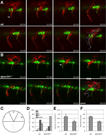
Schwann cells migrate to motor axons in dync1h1 mutant larvae. (A, B) Frames from time-lapse movies of wild-type (A) and dync1h1 mutant (B) larvae carrying Tg(sox10:mRFP) and Tg(mnx1:GFP) transgenes to mark Schwann cells (red, arrow) and motor axons (green). Each sequence starts at 20 hours post fertilization (hpf) and elapsed time is shown in each panel. The final panel in each series shows tracks plotted for the migration of ten RFP+ Schwann cells over 6 hours, with 10 time points per hour. Images shown are lateral views of mid-trunk spinal cord with dorsal up. (C-F) Quantification of Schwann cell progenitor migration. The migration of ten sox10:mRFP+ Schwann cells were tracked over 6 hours with 10 time points per hour. (C) Schwann cell trajectories were assigned a direction of movement based on their starting and ending positions where the apex was assigned as the motor root exit point. I corresponds to anterior to posterior migration, II corresponds to dorsal to ventral migration, and III indicates posterior to anterior Schwann cell migration. A representative track from a wild-type embryo is displayed. (D) Quantification of Schwann cell trajectories from three embryos (30 Schwann cells) indicates that migration to the motor root is reduced but not eliminated in dync1h1 mutants. Categories I, II and III correspond to the diagram in panel C. No MR represents the Schwann cells that did not reach the motor root. (E) Schwann cell velocity is reduced in dync1h1/ embryos (P = 0.004). (F) Schwann cell meandering index, calculated by the displacement from the origin by the track length, is reduced in dync1h1/ embryos (P < 0.0001) indicating less directed movement. P-values were calculated for each track using nonparametric Mann–Whitney U-test statistical analysis. Error bars represent standard error of the mean (SEM). Scale bar equals 40 μm.
|

