
Fig. 2
- ID
- ZDB-FIG-120803-38
- Publication
- McCarroll et al., 2012 - Graded levels of Pax2a and Pax8 regulate cell differentiation during sensory placode formation
- Other Figures
- All Figure Page
- Back to All Figure Page
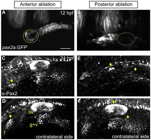
PPA cells are required for normal epibranchial and otic placode development. (A,B) Confocal projection of a Tg(pax2a:GFP)e1 transgenic zebrafish embryo; anterior quarter (A) or posterior third (B) of the PPA was ablated at 12 hpf. Ablated region indicated by dotted line. (C-F) Embryos shown in A and B were Pax2a immunolabeled at 24 hpf, showing expression in otic vesicle and EB placodes. (C,D) Ablated and contralateral sides of the embryo in A. Facial placode on the ablated side exhibits an 81% reduction versus contralateral control; otic and posterior EB placodes are unaffected. (E,F) Ablated and contralateral sides of the embryo in B. Ablated side shows 95% loss of the otic vesicle and 72% reduction in the glossopharyngeal/vagal placodes versus control. Facial placode is unaffected. Arrows indicate corresponding placodes on ablated and control sides. Scale bar: 50 μm |