
Fig. S11
- ID
- ZDB-FIG-120803-22
- Publication
- McCarroll et al., 2012 - Graded levels of Pax2a and Pax8 regulate cell differentiation during sensory placode formation
- Other Figures
- All Figure Page
- Back to All Figure Page
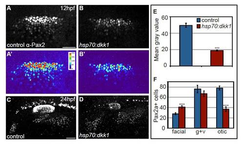
Global Wnt inhibition by Dkk1 expression attenuates Pax2a levels. (A-D) Control and Tg(hsp70:dkk1-GFP)w32 embryos were heat-shocked at 10 hpf and assayed for Pax2a expression at 12 and 24 hpf. Heat maps generated from control (A′) and transgenic embryos (B′) reveal reduced Pax2a levels in the PPA at 12 hpf following heat-shock. Note that upregulation of Dkk1 caused an increase in facial placode size with a concurrent reduction in the size of the otic vesicle. (E) Mean gray values of Pax2a levels in the otic vesicle at 24 hpf revealed a 2.6-fold reduction in the Dkk1-induced embryos (Student’s t-test, ***P<<0.001). (F) Control and Tg(hsp70:dkk1-GFP)w32 embryos were heat-shocked at 10 hpf, collected at 24 hpf and analyzed for Pax2a+ cell number in the otic vesicle and EB placodes. This analysis revealed a 48% increase in the number of cells that segregate to the facial placode (Student’s t-test, ***P<0.001), with a concomitant 47% reduction in the number of cells in the otic placode (Student’s t-test ***P<0.001). There was no significant difference in the number of cells in the glossopharyngeal/vagal placode in these experiments. Scale bar: 50 μm. |