
Fig. 5
- ID
- ZDB-FIG-120803-35
- Publication
- McCarroll et al., 2012 - Graded levels of Pax2a and Pax8 regulate cell differentiation during sensory placode formation
- Other Figures
- All Figure Page
- Back to All Figure Page
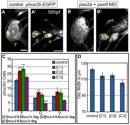
Partial knockdown of pax2a and pax8 transcripts increases cell numbers in EB ganglia. Zebrafish zygotes carrying the TgBAC(phox2b:EGFP)w37 transgene were injected with different amounts of pax2a+pax8 MOs; EB ganglion cell numbers were assayed at 50 hpf. (A,A′) Confocal projection in uninjected control. Facial (F), glossopharyngeal (G) and vagal (V) ganglia (dashed ovals) contain 20, nine and eight EGFP-positive cells, respectively. (B,B2) Confocal projection of an embryo injected with 3+0.25 ng of pax2a+pax8 MOs. Note increased size of G and small V ganglia (F, 29; G, 13; V, 13 cells). (C) Quantification of cells in F, G and small V ganglia in controls and embryos that received increasing doses of pax2a+pax8 MOs. Note significant size increase in all ganglia at [C1]. ***P<0.001, **P<0.005, *P<0.05. (D) Quantification of otic width (longest A-P segment in μm) of controls and embryos receiving increasing doses of pax2a+pax8 MOs (Student′s t-test, P=0.056). n≥33 cells, eight embryos per condition. Error bars represent s.e.m. Scale bars: 10 μm in A; 25 μm in A′. |