
Fig. 4
- ID
- ZDB-FIG-120803-36
- Publication
- McCarroll et al., 2012 - Graded levels of Pax2a and Pax8 regulate cell differentiation during sensory placode formation
- Other Figures
- All Figure Page
- Back to All Figure Page
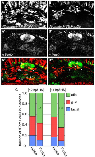
High levels of Pax2a bias otic contribution. (A-B′′) Zebrafish embryos expressing bi-directional heat-shock-inducible plasmid (heat-shocked at 12 and 14 hpf) driving egfp (A-A′′) or pax2a (B-B′′) in one direction and dTomato (dTom) in the other. Arrows indicate Pax2a-misexpressing cells sequestered to otic placode. Arrowheads indicate cells segregated to EB placodes. (C) Relative contribution of dTomato+ cells to the otic vesicle and EB placodes at 24 hpf in EGFP (controls) and Pax2a (overexpression) embryos. Pax2a-overexpressing cells are prone to otic contribution, segregating less frequently to EB placodes versus controls (n≥192 cells from 8-12 embryos per condition; χ2-test; **P<0.016, ***P<0.001). f, facial placode; g+v, glossopharyngeal/vagal placodes; HS, heat-shock; ov, otic vesicle. Scale bars: 50 μm. |