
Fig. 6
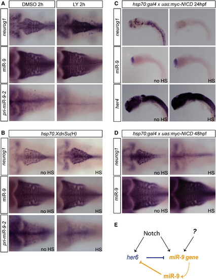
miR-9 Expression Is Regulated by Notch Signaling (A) Expression of neurog1, miR-9, and miR-9-2 in 48 hpf embryos after a 2 h DMSO (left panels) or 10 μM LY411575 treatment (right panels). (B) Expression of neurog1, miR-9, and miR-9-2 in Tg(hsp70:XdnSu(H)) embryos following a heat-shock induction at 48 hpf (HS, right panels) or without any induction (no HS, left panels). (C) Expression of neurog1, miR-9, and her4 in Tg(hsp70l:Gal4)/+;Tg(UAS:myc-NICD)/+ embryos following a heat-shock induction at 24 hpf (HS, right panels) or without any induction (no HS, left panels). (D) Expression of neurog1 and miR-9 in Tg(hsp70l:Gal4)/+;Tg(UAS:myc-NICD)/+ embryos following a heat-shock induction at 48 hpf (HS, right panels) or without any induction (no HS, left panels). (E) Model of regulation of miR-9 by Notch signaling. See also Figure S6. |
| Genes: | |
|---|---|
| Fish: | |
| Conditions: | |
| Anatomical Term: | |
| Stage Range: | Prim-5 to Long-pec |
| Fish: | |
|---|---|
| Conditions: | |
| Observed In: | |
| Stage Range: | Prim-5 to Long-pec |
Reprinted from Developmental Cell, 22(5), Coolen, M., Thieffry, D., Drivenes, O., Becker, T.S., and Bally-Cuif, L., miR-9 Controls the Timing of Neurogenesis through the Direct Inhibition of Antagonistic Factors, 1052-1064, Copyright (2012) with permission from Elsevier. Full text @ Dev. Cell