
Fig. 6
- ID
- ZDB-FIG-120508-49
- Publication
- Zhang et al., 2012 - The Expression of irx7 in the Inner Nuclear Layer of Zebrafish Retina Is Essential for a Proper Retinal Development and Lamination
- Other Figures
- All Figure Page
- Back to All Figure Page
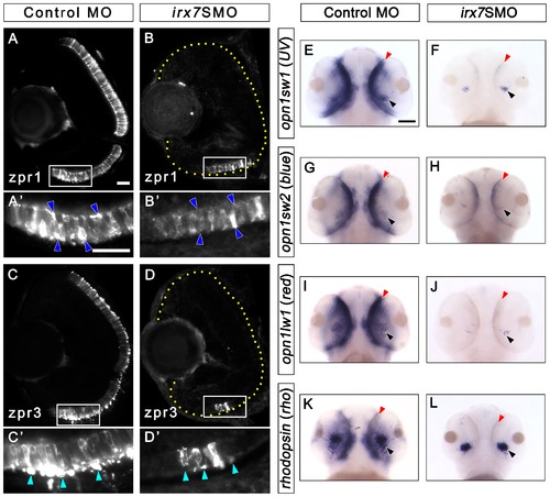
Irx7 knockdown compromises photoreceptor differentiation at 72 hpf. Irx7 knockdown compromised the staining of anti-zpr1 for red-green double cones and anti-zpr3 for rods in the morphants (B and D) compared with the controls at 72 hpf (A and C). (A2, B2, C2 and D2) The corresponding magnified view of the positive signal area in the white box in A, B, C and D. The features indicated by the arrowheads are further discussed in the text. Lateral is to the left and dorsal is up for all sections. In addition, the retinal region in the samples with weak fluorescent signal is highlighted by a dotted yellow line. (E–L) Whole-mount in situ hybridization of three cone opsins (uv, blue and red) and one rod opsin (rho) also indicate the differentiation of these photoreceptors was compromised. The most common staining pattern is shown. The black arrowheads indicate the specific staining (blue colour) of the ventral patch, while the red arrowheads indicate the staining in the ONL. Embryos were imaged from the ventral side and anterior is up. Scale bars = 20 μm for (A)–(D) and 50 μm for (E)–(L). |
| Genes: | |
|---|---|
| Antibodies: | |
| Fish: | |
| Knockdown Reagent: | |
| Anatomical Terms: | |
| Stage: | Protruding-mouth |
| Fish: | |
|---|---|
| Knockdown Reagent: | |
| Observed In: | |
| Stage: | Protruding-mouth |