
Fig. 1
- ID
- ZDB-FIG-120508-44
- Publication
- Zhang et al., 2012 - The Expression of irx7 in the Inner Nuclear Layer of Zebrafish Retina Is Essential for a Proper Retinal Development and Lamination
- Other Figures
- All Figure Page
- Back to All Figure Page
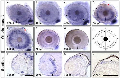
Irx7 is specifically expressed in the prospective INL during zebrafish retinal development. Whole-mount in situ hybridization was conducted to elucidate the expression dynamics of irx7 in embryonic retinas. (A–G) Dissected eyes obtained from embryos between 38 to 96 hpf. Anterior is to the left and dorsal is up. The black arrowheads indicate the irx7+ cells (blue colour) in the retina, the dashed lines indicate the choroid fissure, while the red arrowheads in (D and E) indicate the posterior dorsal region of the retina, the last region to express irx7. (H) A schematic diagram of irx7 expression dynamics in the retina from 38 to 52 hpf. The Roman numerals indicate the order of five retinal regions in which irx7 appears sequentially. (I–L) Transverse retinal section of the corresponding whole-mount embryo at 38, 50, 72 and 96 hpf. Lateral is to the left and dorsal is up. The black arrowheads indicate the irx7+ cells in the retina. Scale bars = 50 μm. |
| Gene: | |
|---|---|
| Fish: | |
| Anatomical Terms: | |
| Stage Range: | Prim-25 to Day 4 |