
Fig. 5
- ID
- ZDB-FIG-120508-48
- Publication
- Zhang et al., 2012 - The Expression of irx7 in the Inner Nuclear Layer of Zebrafish Retina Is Essential for a Proper Retinal Development and Lamination
- Other Figures
- All Figure Page
- Back to All Figure Page
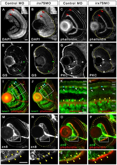
Irx7 knockdown reduces eye size and compromises retinal lamination, INL cells differentiation and dendritic projection of GCs into the INL. Irx7 knockdown reduced eye size and compromised retinal lamination at 72 hpf, as indicated by the DAPI (A and B) and phalloidin (C and D) stains that highlight nuclei and plexiform layers respectively. The red arrows indicate the IPL and OPL. The insets of (A) and (B) also show that the normal elongated morphology of the photoreceptors in control was compromised in the Irx7 morphant. The INL cells differentiation was analyzed by anti-GS for MCs (E and F), anti-PKC for BCs (G and H) and anti-Islet1 for ACs, BCs and HCs (I–L) at the same stage. Irx7 knockdown did not decrease the zn8+ GCs (N) compared with the controls (M), except for the elimination of a fuzzy domain on the apical side of the GCL (compare Q and R). This domain likely represents the dendritic projections of the GCs into the IPL, as it overlapped with the phalloidin staining of the IPL substantially (O and S). This overlap was completely absent in the morphants (P and T). Lateral is to the left and dorsal is up for all sections, except for (K and L), in which the apical side of retina is up. In addition, the retinal region in the samples with weak signal is highlighted by a dotted yellow line. The features indicated by the arrowheads are further discussed in the text. Scale bars = 20 μm. |
| Antibodies: | |
|---|---|
| Fish: | |
| Knockdown Reagent: | |
| Anatomical Terms: | |
| Stage: | Protruding-mouth |