
Fig. S7
- ID
- ZDB-FIG-120202-57
- Publication
- Dohn et al., 2012 - Distinct phases of Wnt/β-catenin signaling direct cardiomyocyte formation in zebrafish
- Other Figures
- All Figure Page
- Back to All Figure Page
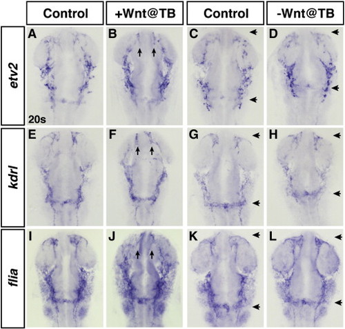
Modulation of Wnt signaling at the TB stage does not significantly affect hemangioblast and endothelial marker genes. (A,C,E,G,I,K) HCSEs. (B,D,F,H,J,L) GFP + embryos. Increased Wnt signaling at the TB stage does not lead to a discernible increase in etv2 (B), a hemangioblast marker, or kdrl (F) or fli1a (J), vascular markers. Arrows in B, F, and J indicate aberrant formation of anterior cerebral veins. (D,H,L) Embryos with decreased Wnt signaling at the TB stage. Decreased Wnt signaling does not cause a discernible decrease in etv2 (D), kdrl (H) or fli1a (L). Although the specification of these lineages does not appear affected, there is a rostral shift of the mandibular arch and endocardial knot (distance between arrows in D,H,L compared to C,G,K). |
Reprinted from Developmental Biology, 361(2), Dohn, T.E., and Waxman, J.S., Distinct phases of Wnt/β-catenin signaling direct cardiomyocyte formation in zebrafish, 364-76, Copyright (2012) with permission from Elsevier. Full text @ Dev. Biol.